Leg Bones And Muscles Diagram / Medical Illustrations Of Superficial Dissection Of Hip And Thigh Showing Lateral Epicondyle And Gerdy S Tubercle - These muscles work together to produce movements such as standing, walking, running, and jumping.
Leg Bones And Muscles Diagram / Medical Illustrations Of Superficial Dissection Of Hip And Thigh Showing Lateral Epicondyle And Gerdy S Tubercle - These muscles work together to produce movements such as standing, walking, running, and jumping.. Human leg muscles diagram human leg muscles. Your leg muscles are some of the hardest working muscles in your body. All four quadriceps muscles insert into the tibia (shin bone). The lower leg extends from the knee to the ankle. See more ideas about muscle anatomy, human anatomy and physiology, body anatomy.
The bones together make up the hip. This area is commonly referred to as the calf. Of course, there are times when it is obvious that the cause of pain is from the bones, joints or skin. The last of the groin muscles is the gracilis, a long, narrow muscle running down the inner thigh like the inseam of a pant leg. The tibia, commonly known as the 'shin bone', is the largest and most medial of the two.you can palpate its anterior border when you run your finger down the anterior aspect of your leg.
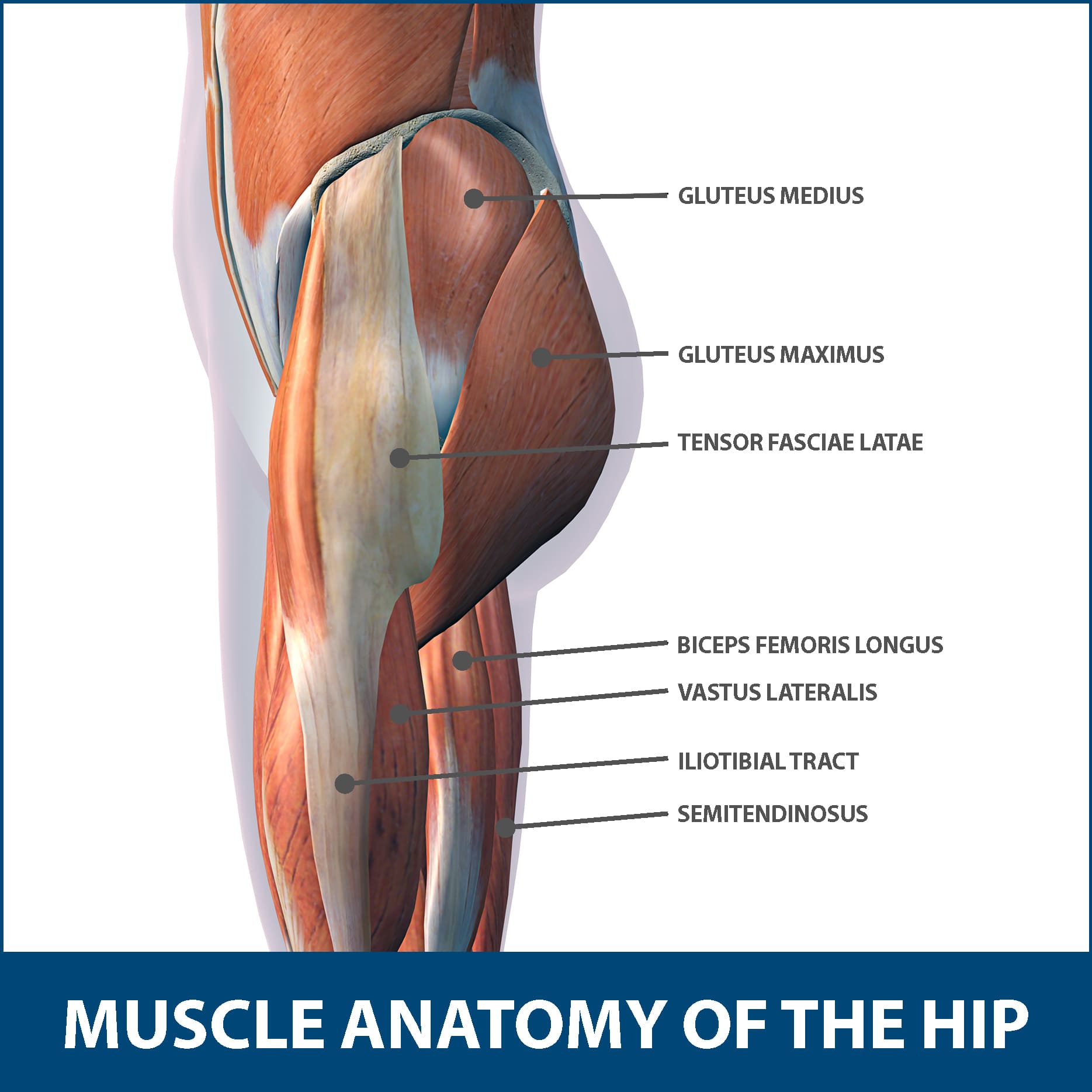
Hip Muscle Strains Info Florida Orthopaedic Institute from www.floridaortho.com Below the gluteus maximus is the smaller gluteus medius. A leg bone is a bone found in the leg. The pain in these conditions is different than muscle pain and there are. The tibia and fibula are two long bones that run parallel to each other, forming the scaffold of the leg and providing attachment points for many muscles. The largest of them is the most superficial muscle, the gluteus maximus. The muscles that pull the legs together, such as those needed when riding a horse, are the adductor muscles of the hip.they originate at the pelvis and attach to the femur. The knee joint is the largest joint in the body and is primarily a hinge joint, although some sliding and rotation occur. It is found on the back of the thigh and runs from the base of the pelvis (specifically the tuberosity of the ischium) to the back of the tibia, one of the bones that make up the lower leg.
Its lower end helps create the knee joint.
3 illustrations of the anterior thigh region detail the anatomy of the femoral quadriceps muscle (rectus femoris, vastus medialis, vastus lateralis and vastus intermedius muscles), the gracilis, sartorius and. Distal end of right humerus. The largest and most medial leg bone, forming both the knee and ankle joints. These muscles work together to produce movements such as standing, walking, running, and jumping. At the same time, the bones and joints of the leg and foot must be strong enough to support the body's weight while remaining. The bones of the leg and foot form part of the appendicular skeleton that supports the many muscles of the lower limbs. The hip itself is a ball and socket joint, much like the shoulder.the structures necessary to create this joint are the socket, the joint capsule, muscle, ligaments, and the neck. A diagram of the pelvis in medial view shows the muscles such as the psoas major, the iliac, piriformis and obturator internus muscles. The pain in these conditions is different than muscle pain and there are. The muscles that pull the legs together, such as those needed when riding a horse, are the adductor muscles of the hip.they originate at the pelvis and attach to the femur. Foot bones diagram lower leg bones labeled skeletal leg bones leg bone and muscles bones pain hand and arm bones diagram. This area is commonly referred to as the calf. Diagram and names of leg bones, diagram of foot and leg bones, diagram of leg bones, diagram of lower leg bones, diagram of the bones in your leg, bone, diagram and.
These muscles work together to produce movements such as standing, walking, running, and jumping. Posted on april 18, 2019april 18, 2019. Leg muscles names leg muscles anatomy human muscle anatomy upper leg muscles leg anatomy anatomy organs thigh muscles gross anatomy abdominal muscles. The bones of the leg and foot form part of the appendicular skeleton that supports the many muscles of the lower limbs. Muscular system medical educational post.
Muscles Of The Hips And Thighs Human Anatomy And Physiology Lab Bsb 141 from s3-us-west-2.amazonaws.com Lower leg muscle diagram blank sketch coloring page. Distal end of right humerus. It is located toward the middle of the lower leg. Use the leg bones diagrams to learn the names of the leg bones. The muscle has several functions, including enabling the leg to flex and rotate. The pain in these conditions is different than muscle pain and there are. The fibula, or calf bone, is smaller and is located on the outside of the lower leg. 3 illustrations of the anterior thigh region detail the anatomy of the femoral quadriceps muscle (rectus femoris, vastus medialis, vastus lateralis and vastus intermedius muscles), the gracilis, sartorius and.
The muscles that pull the legs together, such as those needed when riding a horse, are the adductor muscles of the hip.they originate at the pelvis and attach to the femur.
A superficial muscle, meaning that it lies close to the skin, the gracilis stretches from the pubic symphysis, the joint between the two pubic bones, to the top of the tibia bone in the shin along its medial or inside. Human leg muscles diagram human leg muscles. As these muscles contract and relax, they move skeletal bones to create movement of the body. This chart is beautifully illustrated and offers the most comprehensive look at the muscles of the human leg available. The femur, or thighbone, is the longest and largest bone in the human body. Also, sometimes it is clear that the pain is a result of peripheral neuropathy. Diagram and names of leg bones, diagram of foot and leg bones, diagram of leg bones, diagram of lower leg bones, diagram of the bones in your leg, bone, diagram and. The bones together make up the hip. The muscles that make up the quadriceps are the strongest and leanest of all muscles in the body. 3 illustrations of the anterior thigh region detail the anatomy of the femoral quadriceps muscle (rectus femoris, vastus medialis, vastus lateralis and vastus intermedius muscles), the gracilis, sartorius and. The bones of the hip include the femur, the ilium, the ischium, and the pubis. The lower leg is made up of two very strong, long bone—the tibia and the fibula. A diagram of the pelvis in medial view shows the muscles such as the psoas major, the iliac, piriformis and obturator internus muscles.
It is found on the back of the thigh and runs from the base of the pelvis (specifically the tuberosity of the ischium) to the back of the tibia, one of the bones that make up the lower leg. The largest of them is the most superficial muscle, the gluteus maximus. It is located toward the middle of the lower leg. Diagram and names of leg bones, diagram of foot and leg bones, diagram of leg bones, diagram of lower leg bones, diagram of the bones in your leg, bone, diagram and. Muscular system medical educational post.
Hip Muscle Strains Info Florida Orthopaedic Institute from www.floridaortho.com This muscle runs along the outside of the back of your thigh and attaches to the top of the fibula (the smaller of the two bones of your lower leg). The tibia, also known as the shin bone, is the stronger and larger of the two. Pain in your calf or thigh can be caused by muscle cramps, a pulled or strained muscle, or issues related to your nerves. There are three hamstring muscles, all of them originating at the ischial tuberosity (the bones you sit on): It is found on the back of the thigh and runs from the base of the pelvis (specifically the tuberosity of the ischium) to the back of the tibia, one of the bones that make up the lower leg. Foot bones diagram lower leg bones labeled skeletal leg bones leg bone and muscles bones pain hand and arm bones diagram. The hip itself is a ball and socket joint, much like the shoulder.the structures necessary to create this joint are the socket, the joint capsule, muscle, ligaments, and the neck. The quadriceps muscle attachment points.
It is located toward the middle of the lower leg.
Bones, muscles, joints & ner. Pain in your calf or thigh can be caused by muscle cramps, a pulled or strained muscle, or issues related to your nerves. Use the leg bones diagrams to learn the names of the leg bones. These muscles work together to produce movements such as standing, walking, running, and jumping. Lower leg muscle diagram blank. 3 illustrations of the anterior thigh region detail the anatomy of the femoral quadriceps muscle (rectus femoris, vastus medialis, vastus lateralis and vastus intermedius muscles), the gracilis, sartorius and. This muscle runs along the outside of the back of your thigh and attaches to the top of the fibula (the smaller of the two bones of your lower leg). Most leg pain results from wear and tear, overuse, or injuries in joints or bones or in muscles, ligaments, tendons or other soft tissues. Posted on april 18, 2019april 18, 2019. The muscle has several functions, including enabling the leg to flex and rotate. Distal end of right humerus. The last of the groin muscles is the gracilis, a long, narrow muscle running down the inner thigh like the inseam of a pant leg. Leg muscles names leg muscles anatomy human muscle anatomy upper leg muscles leg anatomy anatomy organs thigh muscles gross anatomy abdominal muscles.
Your leg muscles are some of the hardest working muscles in your body leg bones diagram. Muscular system medical educational post.


0 Komentar