Pacemaker Cells : Excitable Cells - Zoology M with Sheikh at University of ... / Do not interfere with pacemakers, but it is recommended that you keep a cell phone on the opposite side of the body from the pacemaker.
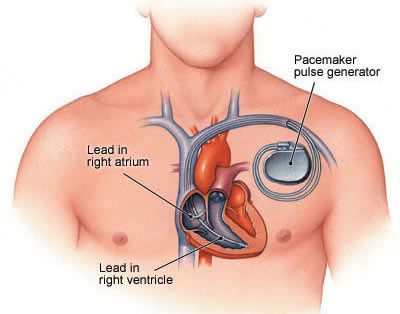
Pacemaker Cells : Excitable Cells - Zoology M with Sheikh at University of ... / Do not interfere with pacemakers, but it is recommended that you keep a cell phone on the opposite side of the body from the pacemaker.. They make up the cardiac pacemaker, that is, the natural pacemaker of the heart. Usually the pacemaker site is the sinoatrial node, near the junction with the superior vena cava. Pacemaker cells cardiac pacemaker cells are mostly found in the sinoatrial (sa) node, which is situated in the upper part of the wall of the right atrium. Implanting a pacemaker in your chest requires a surgical procedure. Rishi is a pediatric infectious disease physic.
Pacemaker cell definition the cells in the sinoatrial node of heart that are involved in creating impulses resulting in the contraction of heart are defined as pacemaker cells. They make up the cardiac pacemaker, that is, the natural pacemaker of the heart. Three different populations of pacemaker cells make up the cardiac conduction system. Pacemaker cells cardiac pacemaker cells are mostly found in the sinoatrial (sa) node, which is situated in the upper part of the wall of the right atrium. The sinoatrial (sa) node or sinus node is the heart's natural pacemaker.
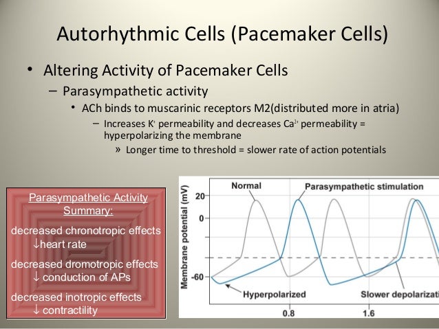
First cardiovascular physiology from image.slidesharecdn.com The sinoatrial (sa) node or sinus node is the heart's natural pacemaker. Myocardial contractile and while myocardial conducting cells. In the pacemaking cells of the heart (e.g., the sinoatrial node ), the pacemaker potential (also called the pacemaker current) is the slow, positive increase in voltage across the cell 's membrane (the membrane potential) that occurs between the end of one action potential and the beginning of the next action potential. A chamber of the heart contracts when an electrical impulse or signal moves across it. There are two major types of cardiac muscle cells: The notion of using a biological pacemaker to replace an electronic pacemaker may be attractive, potentially avoiding the expense and complications associated with device replacement, device or lead failure, and infection; Your surgeon implants it under your skin to help manage irregular heartbeats called arrhythmias. These cells are characterized as having no true resting potential, but instead generate regular, spontaneous action potentials.
In the pacemaking cells of the heart (e.g., the sinoatrial node ), the pacemaker potential (also called the pacemaker current) is the slow, positive increase in voltage across the cell 's membrane (the membrane potential) that occurs between the end of one action potential and the beginning of the next action potential.
The pacemaker cells have an unstable resting membrane potential. When the heart muscle is relaxed the cells are electrically polarized, meaning the inside of each cell has a negative electrical charge. They make up the cardiac pacemaker, that is, the natural pacemaker of the heart. Three different populations of pacemaker cells make up the cardiac conduction system. Modern pacemakers have two parts. Common household devices, such as microwave ovens, tv remotes, heating pads, and electric blankets don't interfere with pacemakers. Pacemaker cells cardiac pacemaker cells are mostly found in the sinoatrial (sa) node, which is situated in the upper part of the wall of the right atrium. Special cells often referred to as pacemaker cells produce electricity in the body by rapidly changing their electrical charge from positive to negative and back again. • more studies are required before the generated cardiac pacemaker cells can be used in the cell therapy. It's used to help your heart beat more regularly if you have an irregular heartbeat (arrhythmia), particularly a slow one. Your surgeon implants it under your skin to help manage irregular heartbeats called arrhythmias. Now, pacemaker cells also listen to which usually come from neighboring pacemaker cells. Cell phones in the u.s.
The collection of pacemaker cells that trigger action potentials in contractile cells; But if those don't come, then a pacemaker cell will simply launch its own and that action potential will then spread around. • generated cardiac pacemaker cells are an excellent testing model for new antiarrhythmics. Rishi is a pediatric infectious disease physic. Cell phones in the u.s.
representation of SAN pacemaker activity. (A) Schematic ... from www.researchgate.net There are two major types of cardiac muscle cells: They are found in all involuntary muscle groups, including both striated and smooth tissues. The cells that create these rhythmic impulses, setting the pace for blood pumping, are called pacemaker cells, and they directly control the heart rate. The action potentials of the pacemaker and contractile cells of the heart differ. We hypothesize that the san contains telocytes that have contacts with pacemaker cells and contractile myocardium. Conduction cells form bundles of fibers that spread the action potential rapidly and sequentially to the contractile myocardium. Special cells often referred to as pacemaker cells produce electricity in the body by rapidly changing their electrical charge from positive to negative and back again. A chamber of the heart contracts when an electrical impulse or signal moves across it.
From cell to bedside (seventh edition), 2018.
Three different populations of pacemaker cells make up the cardiac conduction system. From the nervous system because of the activity of pacemaker cells. It's used to help your heart beat more regularly if you have an irregular heartbeat (arrhythmia), particularly a slow one. Overview of pacemaker cell the heart is composed of two major types of cardiac muscle cells: Do not interfere with pacemakers, but it is recommended that you keep a cell phone on the opposite side of the body from the pacemaker. In the pacemaking cells of the heart (e.g., the sinoatrial node ), the pacemaker potential (also called the pacemaker current) is the slow, positive increase in voltage across the cell 's membrane (the membrane potential) that occurs between the end of one action potential and the beginning of the next action potential. The cells that create these rhythmic impulses, setting the pace for blood pumping, are called pacemaker cells, and they directly control the heart rate. When the heart muscle is relaxed the cells are electrically polarized, meaning the inside of each cell has a negative electrical charge. Pacemaker cell definition the cells in the sinoatrial node of heart that are involved in creating impulses resulting in the contraction of heart are defined as pacemaker cells. But if those don't come, then a pacemaker cell will simply launch its own and that action potential will then spread around. This is called automaticity, and that's easy to remember because it's got automatic right in it. Cardiac pacemaker a small mass of specialized muscle tissue in the heart that sets a rhythm of contraction and relaxation for the other parts of the heart, resulting in the heartbeat. Myocardial contractile and while myocardial conducting cells.
The action potentials of the pacemaker and contractile cells of the heart differ. This is called automaticity, and that's easy to remember because it's got automatic right in it. The collection of pacemaker cells that trigger action potentials in contractile cells; Common household devices, such as microwave ovens, tv remotes, heating pads, and electric blankets don't interfere with pacemakers. The myocardial contractile cells constitute the bulk (99 percent) of the cells in the atria and ventricles.
What Is the Pacemaker of the Heart? | New Health Guide from www.newhealthguide.org From cell to bedside (seventh edition), 2018. Cell phones in the u.s. These cells have natural automaticity, meaning they can generate their own action potentials. The notion of using a biological pacemaker to replace an electronic pacemaker may be attractive, potentially avoiding the expense and complications associated with device replacement, device or lead failure, and infection; These cells are characterized as having no true resting potential, but instead generate regular, spontaneous action potentials. Pacemaker cells cardiac pacemaker cells are mostly found in the sinoatrial (sa) node, which is situated in the upper part of the wall of the right atrium. From the nervous system because of the activity of pacemaker cells. In the pacemaking cells of the heart (e.g., the sinoatrial node ), the pacemaker potential (also called the pacemaker current) is the slow, positive increase in voltage across the cell 's membrane (the membrane potential) that occurs between the end of one action potential and the beginning of the next action potential.
From cell to bedside (seventh edition), 2018.
The collection of pacemaker cells that trigger action potentials in contractile cells; We hypothesize that the san contains telocytes that have contacts with pacemaker cells and contractile myocardium. Find out how the pacemaker cells use the movement of sodium, calcium, and potassium to get your heart beating! Adapted to continued rhythmic contraction Usually the pacemaker site is the sinoatrial node, near the junction with the superior vena cava. A pacemaker is a small device that sends electrical impulses to the heart muscle to maintain a suitable heart rate and rhythm. The action potentials of the pacemaker and contractile cells of the heart differ. A pacemaker is an electrically charged medical device. Cells within the sinoatrial (sa) node are the primary pacemaker site within the heart. Now, pacemaker cells also listen to which usually come from neighboring pacemaker cells. It produces the electrical impulses that cause your heart to beat. These cells are characterized as having no true resting potential, but instead generate regular, spontaneous action potentials. Your surgeon implants it under your skin to help manage irregular heartbeats called arrhythmias.
This is called automaticity, and that's easy to remember because it's got automatic right in it pacemaker. There are two major types of cardiac muscle cells:


0 Komentar