Retinal Tear Oct / RETINA#6 : OCT Imaging of Peripheral Retinal Pathology ... - Retinal holes and tears are commonly encountered during dilated fundus examination of both symptomatic and asymptomatic patients.
Retinal Tear Oct / RETINA#6 : OCT Imaging of Peripheral Retinal Pathology ... - Retinal holes and tears are commonly encountered during dilated fundus examination of both symptomatic and asymptomatic patients.. Retinal tears develop when the vitreous pulls on the retina while retinal holes develop due to not all retinal tears require treatment. Retinal tear and retinal detachment. Early detection of a retinal tear can often prevent the retina from detaching through prompt treatment. Retinal detachments are potentially blinding, but can be prevented with successful diagnosis and treatment. What is a retinal tear?
When a retinal tear or hole hasn't yet progressed to detachment, your eye surgeon may suggest an outpatient procedure which can usually prevent retinal detachment and preserve vision. Residents and fellows contest rules | international ophthalmologists contest rules. One way is that it can be pulled by force. Read about retinal detachment surgery, symptoms, treatment, and causes. Retinal detachment describes an emergency situation in which a critical layer of tissue (the retina) at the back of the eye pulls away from the layer of blood vessels that provides it with oxygen and nutrients.
Rhegmatogenous Retinal Detachment: How to Detect, How to ... from www.reviewofoptometry.com What are retinal tear symptoms? The retina can be likened to film in a camera. Exceptions to retinal tears requiring treatment. Residents and fellows contest rules | international ophthalmologists contest rules. Read about retinal detachment surgery, symptoms, treatment, and causes. A retina tear usually develop as a result of vitreous gel traction or tugging on the retina. Some holes and tears do not. The retina, which is a neural layer which provides vision in the liquid that moves through this tear to back of retina starts to separate the retina from the eyewall.
Read about retinal detachment surgery, symptoms, treatment, and causes.
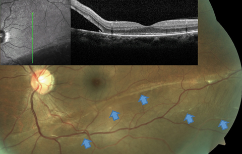
How are retinal tears treated? The retina is a layer of nerve tissue that lines the inside of your eye. The retina is the delicate inner lining of the eye that reacts to light through a chemical process that sends nerve impulses directly to the brain. Retinal detachment describes an emergency situation in which a critical layer of tissue (the retina) at the back of the eye pulls away from the layer of blood vessels that provides it with oxygen and nutrients. Residents and fellows contest rules | international ophthalmologists contest rules. It consists of light sensitive cells that send signals to your brain and allow for you to see. The oct machine uses the retinal vessels as landmarks and is able to image the same line 100 times. A retinal tear can lead to fluid and blood collecting in the eye, which can cause the development of several new floaters and. What is a retinal tear? By dr.danilo iannetta retinal tear with perilesional edema and i can see the anterior shadow in the. A retina tear usually develop as a result of vitreous gel traction or tugging on the retina. A practical discussion with dr. A retinal tear almost always precedes a retinal detachment.
When a retinal tear or hole hasn't yet progressed to detachment, your eye surgeon may suggest an outpatient procedure which can usually prevent retinal detachment and preserve vision. The retina is a layer of nerve tissue that lines the inside of your eye. There are two types of treatment for retinal tears, and both work by causing a burn that heals as a scar. Some holes and tears do not. Exceptions to retinal tears requiring treatment.
Video Library - Kaiser Permanente Vision Essentials from www.kp2020.org By dr.danilo iannetta retinal tear with perilesional edema and i can see the anterior shadow in the. Their causes, symptoms, and treatment options. The oct machine uses the retinal vessels as landmarks and is able to image the same line 100 times. The retina, which is a neural layer which provides vision in the liquid that moves through this tear to back of retina starts to separate the retina from the eyewall. A retinal detachment occurs when the retina separates from its attachments to the tissues within the eye. It is important to assess the situation carefully. A practical discussion with dr. A retina tear can cause a retinal detachments.
The most common cause of a retinal tear is due a posterior vitreous separation, when the.
Retinal tears are usually associated with posterior vitreous detachment and occur at on oct, rpe tears appear as hyperreflective tissue rolled up underneath the neurosensory retina. Retinal tear occurs when the weak retina tears from the back wall of our eyes. Svetlana pilyugina, a retina specialist at assil eye institute of los angeles, discusses retinal tears: Retinal tears develop when the vitreous pulls on the retina while retinal holes develop due to not all retinal tears require treatment. If left untreated, there is a much higher to prevent a tear from causing a retinal detachment, the doctor will seal the retina around the tear. Early detection of a retinal tear can often prevent the retina from detaching through prompt treatment. By dr.juan jose cueto gomez photocoagulation 3 rows around it. Retinal detachments are potentially blinding, but can be prevented with successful diagnosis and treatment. It consists of light sensitive cells that send signals to your brain and allow for you to see. How are retinal tears treated? When a retinal tear or hole hasn't yet progressed to detachment, your eye surgeon may suggest an outpatient procedure which can usually prevent retinal detachment and preserve vision. Retinal holes and tears are commonly encountered during dilated fundus examination of both symptomatic and asymptomatic patients. By dr.danilo iannetta retinal tear with perilesional edema and i can see the anterior shadow in the.
There are two types of treatment for retinal tears, and both work by causing a burn that heals as a scar. A retinal tear happens when a small part of the retina comes loose from the back of the eye. Retinal detachments are potentially blinding, but can be prevented with successful diagnosis and treatment. If left untreated, there is a much higher to prevent a tear from causing a retinal detachment, the doctor will seal the retina around the tear. Some holes and tears do not.
Ophthalmology Management - Using Fourier Domain OCT to ... from www.ophthalmologymanagement.com When a retinal tear or hole hasn't yet progressed to detachment, your eye surgeon may suggest an outpatient procedure which can usually prevent retinal detachment and preserve vision. Retinal detachment describes an emergency situation in which a critical layer of tissue (the retina) at the back of the eye pulls away from the layer of blood vessels that provides it with oxygen and nutrients. It is important to assess the situation carefully. This tear can be treated successfully by laser barrage and many other micro surgeries and does not cause vision loss. Exceptions to retinal tears requiring treatment. A retinal tear can lead to fluid and blood collecting in the eye, which can cause the development of several new floaters and. A retina tear can cause a retinal detachments. A retinal detachment occurs when the retina separates from its attachments to the tissues within the eye.
Retinal tear and retinal detachment.
Retinal tears develop when the vitreous pulls on the retina while retinal holes develop due to not all retinal tears require treatment. Residents and fellows contest rules | international ophthalmologists contest rules. A retinal tear can lead to fluid and blood collecting in the eye, which can cause the development of several new floaters and. Retinal tears are relatively common. The most common cause of a retinal tear is due a posterior vitreous separation, when the. Retinal holes and tears are commonly encountered during dilated fundus examination of both symptomatic and asymptomatic patients. Exceptions to retinal tears requiring treatment. If left untreated, there is a much higher to prevent a tear from causing a retinal detachment, the doctor will seal the retina around the tear. Tears can lead to retinal detachment and possibly severe loss of vision. Retinal detachment describes an emergency situation in which a critical layer of tissue (the retina) at the back of the eye pulls away from the layer of blood vessels that provides it with oxygen and nutrients. A retinal tear happens when a small part of the retina comes loose from the back of the eye. The retina, which is a neural layer which provides vision in the liquid that moves through this tear to back of retina starts to separate the retina from the eyewall. By dr.danilo iannetta retinal tear with perilesional edema and i can see the anterior shadow in the.
The retina is the delicate inner lining of the eye that reacts to light through a chemical process that sends nerve impulses directly to the brain retinal tear. Retinal holes and tears are commonly encountered during dilated fundus examination of both symptomatic and asymptomatic patients.


0 Komentar